Diseases caused by fungal infections
Pathogenesis of fungal infections
- Opportunistic growth
- Tissue damage
- Virulence factors
- Capsule (Cryptococcus)
- Keratinase
- Inflammatory immune response
Host defenses
- Primarily cell-mediated
- TH1 - cytokines
- Inflammation
- IFN-gamma: activate macrophages
Antifungal treatment
- Topical
- Systemic: amphothericin B, fluorocytosine
Types of fungal infections
- Superficial: Hair and skin surface
- Cutaneous: keratinized tissue
- Subcutaneous: more invasive
- Systemic
Diseases caused by fungi
- Superficial mycoses
- Growth of fungi on surface of skin or hair
- Clinical conditions
- Cutaneous mycoses
- Often called ringworm (Tinea)
- Typically caused by 3 groups of molds
- Microsporum
- Epidermophyton
- Trichophyton
- May be carried by humans, animals or found in soil
- Subcutaneous mycoses
- More invasive and destructive
- Elicit immune reaction
- Sometimes difficult to treat; may require surgery or amputation
- Clinical diseases
- Sporotrichosis
- Cause: Sporothrix schenckii
- Trauma/wound entry of spores
- Ulcering lesions along lymphatic duct affected
- Chromoblastomycosis
- Cause: various pigment-producing fungi
- Warty, cauliflower-like skin lesions
- Eumycotic mycetoma
-
Subcutaneous zygomycosis
- Conidiobolus and Basidiobolus
- Extensive swelling in affected area
- Subcutaneous Phaeohyphomycosis
- Dematiacious fungi (e.g. Phialophora, Exophiala)
- single cyst; may expand to cover more body surfaces
- systemic infection occurs in immunocompromised patients
- Systemic mycoses
- Caused by virulent fungi
- Can cause disease in healthy individuals
- Can evade host defenses
-
Many of these fungi are dimorphic
- Mold phase found in nature
- Yeast phase is pathogenic stage
- Clinical Diseases
- Cause: Histoplasma capsulatum
- Endemic in Midwestern US
- Associated with bats and birds
- Pneumonia in 5% of infected
- Disseminated progressive disease (immunocompromised)
- Cause: Blatomyces dermatitidis
- Endemic in North America
- Associated with dogs
- Primary infection is usually asymptomatic
- Blastomyces can survive in macrophages = dissemination
- Pulmonary disease
- Disseminated disease with bone and brain lesions
- Cause: Coccidioides immitis
- Endemic in the Americas
- Associated with arid soil
- ~ 40% of infections are symptomatic: benign respiratory
- Progressive disease in lungs
- Systemic disease involves skin & meninges
- Cause; Paracoccidioides braziliensis
- Dimorphic yeast/mold
- Limited to South America
- Inhalation of spores
- Usually self-limiting, but disease may remain dormant and reactivate
later
- Infection can spread to multiple tissues and organs
Opportunistic mycoses
- Cause: Cryptococcus neoformans
- Encapsulated yeast
- Associated with pigeons
- Fungal meningitis & disseminated disease in AIDS
- Lung infection leads to nodule formation
- Meningitis (headache, fever, mental changes)
- Skin and bone lesions
- Causes: Candida albicans et al.
- Primarily a problem in the compromised host
- Wounds
- Antimicrobial drugs
- Immunosuppression
- AIDS
- External candidiasis: Thrush, vaginosis
- Chronic candidiasis: Drug-resistant mucocutaneous candida
- Systemic candidiasis: various organs, incl. brain
- not a big problem in AIDS
- Cause: Aspergillus flavus and A. fumigatus
- Food intoxication (aflatoxin)
- Hypersensitivity pulmonary disease (allergic reaction)
- Colonization
- Systemic disease (invasive)
- Hyphal growth in blood vessels and tissues
- Blockage of blood vessels by hyphae and clots
- Tissue necrosis
- Cause: Rhizopus, Mucor
- Rhinocerebral zygomycosis: sinuses, eyes, palate, brain
- terminally acidotic/diabetic patients
- Colonization of other organs and tissues
- Dissemination with growth in major blood vessels
- Cause: Pneumocystis jirovecii
- Common in old age, immunosuppression, and AIDS
- Common in human population (asymptomatic) and rats
- Causes interstitial pneumonitis, fever, cyanosis
- Resistant cysts formed
Unusual infections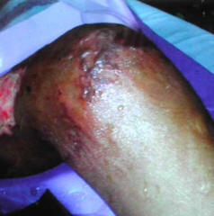
- Adiaspiromycosis (Fig. 1)
- Emmonsia spp.
- causes granulomas in lungs
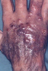
- Lobomycosis (Fig. 2)
- Chronic cutaneous infection (nodules) caused by Lacazia loboi
- South/central Americas; also found in dolphins
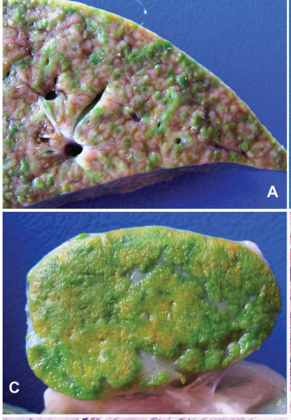
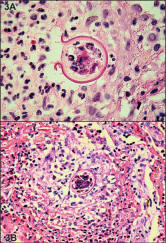
- Chlorellosis (Fig. 3)
- Caused by green algae
- usually sheep/cattle infection
- Protothecosis (Fig. 4)
- chloroplast-less algae (Prototheca wickerhamii)
- skin nodules; usually in immunocompromised
- Pythiosis insidiosis (Fig. 5)
- Caused by protist microbe Pythium insidiosis (oomycete)
- Ischemia and tissue necrosis in lower limbs; usually a veterinary
disease