
Laboratory Investigations in Microbiology
|
Laboratory Investigations in Microbiology |
|
Although respiration, be it aerobic or anaerobic, is the most efficient form of energy generation, not all bacteria can do respiration at all times. A less efficient alternative, called fermentation, is found in many bacteria. Some, such as Streptococcus, are entirely fermentative and do not have the ability to do respiration. Other bacteria choose fermentation when the electron acceptors of respiration are unavailable. Fermentation generates only ~10% of the energy that can be gained by respiration (per molecule of food used). In part, cells make up for this difference by fermenting significantly larger quantities of food than they would use in respiration alone. The advantage of fermentation, however, is that no external electron acceptor (such as oxygen or nitrate) is needed. The most common electron acceptor in fermentation is pyruvate, produced through glycolysis. Thus, bacteria that break down glucose to pyruvate can turn around and immediately use this pyruvate as an electron acceptor. The bacteria keep only the ATP generated during glycolysis and the reduced form of pyruvate (often lactic acid) is lost to the cell as a waste product.
Fermentation is usually carried out in anaerobic environments, since facultative anaerobes will resort to respiration if oxygen is present. As bacteria ferment their food source, the fermentation products are released into the culture medium. Such fermentation products include organic acids (lactate, butyrate, acetate, formate, propionate), alcohols (ethanol, isopropanol, butanol, butanediol), and gases (CO2, H2). Because bacteria produce large quantities of fermentation byproducts, the culture medium is changed. By using a few simple indicators (such as phenol red, a pH indicator), we can detect the formation of acids, gases, and even butanediol. This forms the basis of the fermentation tests we will conduct today.
 Glucose
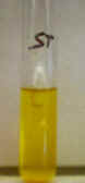
fermentation test (Atlas p. 52): Fermentation of glucose, one of the most easily
used carbohydrates, is tested using a fermentation broth containing peptone
(protein), glucose, phenol red (a pH indicator), and a Durham tube (an
upside-down small glass tube inside the broth). When acid is produced, the
color of the medium changes from red/orange (neutral) to yellow. This
indicates a positive test for acid production. This test does not tell
us which acids are made or how much acid is produced. An orange or
red color
indicates a negative test for fermentation.
Glucose
fermentation test (Atlas p. 52): Fermentation of glucose, one of the most easily
used carbohydrates, is tested using a fermentation broth containing peptone
(protein), glucose, phenol red (a pH indicator), and a Durham tube (an
upside-down small glass tube inside the broth). When acid is produced, the
color of the medium changes from red/orange (neutral) to yellow. This
indicates a positive test for acid production. This test does not tell
us which acids are made or how much acid is produced. An orange or
red color
indicates a negative test for fermentation.
Some bacteria attack proteins in the medium if they cannot do fermentation. When they do, amino acids are broken down, releasing ammonium (see chapter 12). As a result, the medium turns magenta in color, indicating base production.
When gas (CO2 or H2) is produced from fermentation, bubbles will be trapped inside the Durham tube. This indicates a positive test for gas production.
Production of alcohol (ethanol, e.g.) cannot be detected with this test.
Sucrose and lactose fermentation tests:
These two tests work the same way as the glucose test. Instead of glucose,
however, the disaccharides lactose or sucrose are used. 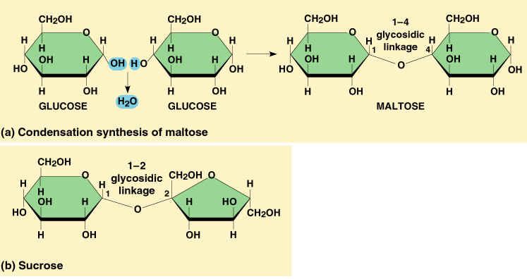 Only bacteria that
have the enzymes to hydrolyze these disaccharides first (producing glucose +
one other monosaccharide) can ferment them! For these tests to be positive,
bacteria must be able to do fermentation (see glucose test) AND have the
enzyme to break down the disaccharide. A negative test in this case can
have two explanations: the bacteria do not carry the disaccharidase necessary,
or the bacteria do not carry out fermentation. Positive fermentation results are
scored the same way as in the glucose test.
Only bacteria that
have the enzymes to hydrolyze these disaccharides first (producing glucose +
one other monosaccharide) can ferment them! For these tests to be positive,
bacteria must be able to do fermentation (see glucose test) AND have the
enzyme to break down the disaccharide. A negative test in this case can
have two explanations: the bacteria do not carry the disaccharidase necessary,
or the bacteria do not carry out fermentation. Positive fermentation results are
scored the same way as in the glucose test.
GLS combination test. Many bacterial identification systems available commercially use multiple test chambers/wells to test for the ability of a microbe to use or ferment a variety of sugars, and the overall pattern of sugar use can be used to identify the microbe. The GLS test is a scaled-down version using only 3 sugars (Glucose, Lactose, Sucrose), but even this milited combination allows for 5 different outcomes. This allows a more rapid distinction among test microbes. The main limitation in this test is that it only detects Acid (A) so the outcome for each sugar is evaluated as either positive (A) or negative (-). No gas can be detected.
Voges Proskauer (VP) test (Atlas p. 82):
Although traditional fermentatio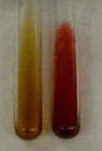 n
tests can detect acids and gases, the precise fermentation
pathway followed cannot be deduced. Some bacteria produce only lactic acid (homolactic
fermentation), while others produce a mixture of acids and gases (mixed
acid fermentation) or acids, gases, and butanediol (butanediol
fermentation). To distinguish some of these possibilities, one can test for
specific chemicals, such as acetoin (the precursor for butanediol). The Voges
Proskauer test identifies bacteria that produce butanediol. The reagents (phenol
+ NaOH) react with acetoin, a butanediol precursor, to produce a red color. This
indicates a positive VP test. In a negative VP test, the reagents may
be brown, yellow, or green in color.
n
tests can detect acids and gases, the precise fermentation
pathway followed cannot be deduced. Some bacteria produce only lactic acid (homolactic
fermentation), while others produce a mixture of acids and gases (mixed
acid fermentation) or acids, gases, and butanediol (butanediol
fermentation). To distinguish some of these possibilities, one can test for
specific chemicals, such as acetoin (the precursor for butanediol). The Voges
Proskauer test identifies bacteria that produce butanediol. The reagents (phenol
+ NaOH) react with acetoin, a butanediol precursor, to produce a red color. This
indicates a positive VP test. In a negative VP test, the reagents may
be brown, yellow, or green in color.
Methyl Red (MR) test (Atlas p. 63): 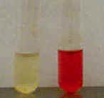 One
can also determine the amount of acid produced using the MR test. Methyl
red is a pH indicator that turns from yellow (neutral) to red (acid) at a pH of
~ 4.4. bacteria that carry out Mixed acid fermentation produce many strong acids
(lactic, acetic, formic) that lower the pH below 4.4, resulting in a red
color when the methyl red is added. This indicates a positive MR
test. Other bacteria produce weaker and/or fewer acids and therefore do not
lower the pH of the medium as much, resulting in an orange or yellow color when
methyl red is added. This indicates a negative MR test. Please note
that these color changes are opposite of those in the phenol red test!
One
can also determine the amount of acid produced using the MR test. Methyl
red is a pH indicator that turns from yellow (neutral) to red (acid) at a pH of
~ 4.4. bacteria that carry out Mixed acid fermentation produce many strong acids
(lactic, acetic, formic) that lower the pH below 4.4, resulting in a red
color when the methyl red is added. This indicates a positive MR
test. Other bacteria produce weaker and/or fewer acids and therefore do not
lower the pH of the medium as much, resulting in an orange or yellow color when
methyl red is added. This indicates a negative MR test. Please note
that these color changes are opposite of those in the phenol red test!
After 24 - 48 hours:
After 24 - 48 hours:
After 24 - 48 hours:
© 2003 - 2019 José de Ondarza, Ph.D.