
Laboratory Investigations in Microbiology
|
Laboratory Investigations in Microbiology |
|
Differential stains can distinguish bacteria based on their affinity for certain dyes and the ease with which cells can be de-stained. The two differential stains we will observe here are the Gram stain and the acid-fast stain.
One of the most commonly used staining procedures in Microbiology is the Gram Stain (Atlas p. 35 - 38). Named after Christian Gram, this procedure distinguishes among two types of bacteria: Gram-positive and Gram-negative (Atlas Fig. 5.2). Because this stain makes a distinction between bacteria, it is known as a differential stain. The Gram stain procedure involves four major steps (Atlas Fig. 5.1):
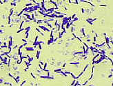 Gram-positive
cells are turned purple with the crystal violet. The iodine fixes the
stain in the cells so that it does not easily wash out, and the alcohol acts on
the thick peptidoglycan cell wall of Gram-positive
cells in such a way as to trap the dye in the cells rather than
washing it away. Hence, Gram-positive cells stay
purple. The safranin also enters the cells, but the darker crystal violet
masks it.
Gram-positive
cells are turned purple with the crystal violet. The iodine fixes the
stain in the cells so that it does not easily wash out, and the alcohol acts on
the thick peptidoglycan cell wall of Gram-positive
cells in such a way as to trap the dye in the cells rather than
washing it away. Hence, Gram-positive cells stay
purple. The safranin also enters the cells, but the darker crystal violet
masks it. 
Gram-negative cells also are turned purple by the crystal violet, but the iodine mordant is not sufficient to retain the dye in the cells when they are rinsed with alcohol. Since Gram-negative cells have a very thin layer of peptidoglycan, the crystal violet is rinsed out, and the cells become colorless. Counterstaining with safranin then turns Gram-negative cells pink.
It is, however, important to note that not all bacteria follow this pattern. Some Gram-negative cells (e.g. Deinococcus), when stained, appear purple because they have an unusual cell wall structure that retains the crystal violet like Gram-positive cells do. Some Gram-positive cells may, in an older culture, stain pink or pink + purple ("Gram-variable") even though they have a regular Gram-positive wall. Lastly, the meaning of Gram-positive and Gram-negative is lost when applying this technique to Archaea or eukaryotes - since these organisms do not have peptidoglycan cell walls! Nonetheless, this staining procedure can be used as an identifying trait of virtually any bacterial species.
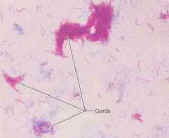 The Acid-fast stain is used to identify
bacteria such as Mycobacterium tuberculosis. Mycobacteria have a
tough waxy cell wall that makes them tough to kill. This cell wall also
resists destaining with acids, and therefore this procedure can distinguish
acid-fast bacteria (which keep the dye) from other bacteria (which become
colorless).
The Acid-fast stain is used to identify
bacteria such as Mycobacterium tuberculosis. Mycobacteria have a
tough waxy cell wall that makes them tough to kill. This cell wall also
resists destaining with acids, and therefore this procedure can distinguish
acid-fast bacteria (which keep the dye) from other bacteria (which become
colorless).
The goals of today's lab exercise are to perform a successful Gram stain and to use this procedure to distinguish various types of 'unknown' bacteria.
© 2003 - 2015 José de Ondarza, Ph.D.