
Laboratory Investigations in Microbiology
|
Laboratory Investigations in Microbiology |
|
Bacteria have several means by which they can move through their environment. By far the most common is flagellar motility, but other forms include gliding, sliding and twitching movement.
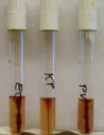
Motility deeps. For bacteria that are motile via flagella, a simple test for motility is the motility deep. This agar deep has a low concentration of agar to allow motile bacteria to move through it, while non-motile bacteria are kept in the stab line. This test is like the motility test using the SIM deep. However, this agar medium is clear in color and contains a dye that turns pink in the presence of bacteria. Hence, you can see exactly where in the test tube the bacteria have gone (or stayed). In this picture on the right, PV and EC show motility, whereas KP remains within the stab line and is non-motile.
The key to interpretation of bacterial motility is to look for a pink color that spreads away from the stab line into the agar around it. The agar medium around the stab line should be completely transparent for non-motile bacteria. Be sure, however, not to confuse dense growth within the stab line for motility.
One note of caution; this test does not work well for all bacteria. Most Gram-positive bacteria, even if motile, form chains or clumps and do not move well into the agar medium. Likewise, strictly aerobic bacteria grow mainly on the surface of the agar deep and do not grow well inside the deep, making it hard to determine motility. Lastly, bacteria which do not reduce the dye in the medium will not evidence a good coloration. Therefore this test is best suited for distinguishing motility in facultatively anaerobic Gram-negative rods, such as the Enterobacteriaceae.
Darkfield or phase contrast microscopy. Bacterial motility can also be determined by direct observation under phase contrast or dark field illumination. Live, unstained bacteria are viewed under 400 - 1000x magnification. Motility is easily observable, but note the difference between the rapid swimming motility (flagellar) seen in E. coli and the random movements produced by Brownian motion in non-motile bacteria such as Staphylococcus (see video link below).
Next lab period:
© 2003 - 2017 José de Ondarza, Ph.D.