
Laboratory Investigations in Microbiology
|
Laboratory Investigations in Microbiology |
|
Bacterial growth on various culture media
Bacterial growth patterns depend on the type of medium they are in. Broth cultures go through the four stages of microbial growth sequentially: lag phase, exponential phase, stationary phase, and death phase. Because growth of bacteria in broths turns the broth cloudy, the extent of growth can be measured by spectrophotometry (Ch. 9). Observe some broth cultures, and you will also be able to discern a difference in the way various cultures grow in broths.
 Bacterial
growth on agar surfaces results in the formation of bacterial colonies. A
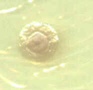
bacterial colony is a visible accumulation of bacterial cells that arise from
one single cell. Therefore, all cells in a pure colony are genetically identical
(clones). Notice that bacterial colonies are not always uniform. The center of a
colony often consists of cells in stationary or death phase, while the cells at
the edges of a colony are actively dividing (exponential phase). This often
causes the center of a colony to look different from its edges.
Bacterial
growth on agar surfaces results in the formation of bacterial colonies. A
bacterial colony is a visible accumulation of bacterial cells that arise from
one single cell. Therefore, all cells in a pure colony are genetically identical
(clones). Notice that bacterial colonies are not always uniform. The center of a
colony often consists of cells in stationary or death phase, while the cells at
the edges of a colony are actively dividing (exponential phase). This often
causes the center of a colony to look different from its edges.
As we will soon see, microbial growth is affected by many factors, including temperature, pH, oxygen, and nutrient availability. When optimally adjusted, these growth conditions can support rapid bacterial growth. The following lab exercise will allow us to observe bacterial growth on various culture media and to practice the techniques for obtaining isolated colonies and preparing pure cultures.
Culture techniques
Streak-plate technique: The most
frequently used method for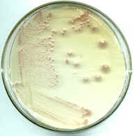 obtaining colonies of bacteria on agar surfaces is
the streak plate technique. The photo on the right shows a culture of Serratia
marcescens spread across an agar plate using this technique. On the bottom left
is the first streak, made with a loopful of broth culture. A second streak is
made with a sterile loop that crosses into the first streak, spreading some of
the bacteria out more (top left). A third (and sometimes fourth) streak spreads
those bacteria even more, until in the end single bacterial cells are spread
out. After incubation, the bacteria form colonies - visible piles of bacterial
cells. Notice that the colonies that are most isolated are also the largest. Why
do you think this is so? {Answer} You can also see
that these bacterial colonies produce a red pigment. Colony characteristics such
as color, shape, size, and margin can be useful in identifying a bacterial
species.
obtaining colonies of bacteria on agar surfaces is
the streak plate technique. The photo on the right shows a culture of Serratia
marcescens spread across an agar plate using this technique. On the bottom left
is the first streak, made with a loopful of broth culture. A second streak is
made with a sterile loop that crosses into the first streak, spreading some of
the bacteria out more (top left). A third (and sometimes fourth) streak spreads
those bacteria even more, until in the end single bacterial cells are spread
out. After incubation, the bacteria form colonies - visible piles of bacterial
cells. Notice that the colonies that are most isolated are also the largest. Why
do you think this is so? {Answer} You can also see
that these bacterial colonies produce a red pigment. Colony characteristics such
as color, shape, size, and margin can be useful in identifying a bacterial
species.
Spread-plate technique: At times it is convenient to merely spread a sample of bacteria out on an agar plate using a sterile glass rod. This works well if the number of bacteria in a sample is relatively small - maybe 100 - 1000 cells. Spread plates distribute bacteria more or less equally across the agar surface, and more than 1000 colonies would make it difficult to isolate pure colonies.
Pour-plate technique: This technique is useful when bacterial numbers are to be determined. Instead of placing bacteria on top of the agar, they are mixed with the agar medium while it is still melted (~ 50C). Immediately thereafter, the mixture is poured into a sterile Petri plate where it hardens. Most of the bacteria will be embedded in the agar and will form smaller, denser colonies. This makes it possible to count several hundred colonies on one plate. However, it is more difficult to isolate pure strains and to observe the true colony characteristics. This technique will be demonstrated at a later time.
Preparation ![]() of
pure cultures: Using a streak plate, spread plate, or contamination
plate, pure cultures of each of the bacteria or fungi on the plate can be
prepared if isolated colonies of the bacteria can be found. A isolated colony is
one that is visibly uncontaminated and does not come in contact with any other
colonies. In order to transfer bacteria from an isolated colony, aseptic technique is used. The tip of
an inoculating loop is dipped into the colony (you don't need much of a colony -
keep in mind that millions of cells can be clinging to a loop without you even
seeing them), then
placed onto the agar slant surface near the bottom of the test tube and
gradually pulled out of the tube. By wiggling the loop back and forth as it is
being withdrawn, a curvy line is formed on the agar surface. {I liken this to
skiing down a ski slope - from the top of the slope (in the bottom of the tube)
to the end (near the mouth of the test tube)}.
of
pure cultures: Using a streak plate, spread plate, or contamination
plate, pure cultures of each of the bacteria or fungi on the plate can be
prepared if isolated colonies of the bacteria can be found. A isolated colony is
one that is visibly uncontaminated and does not come in contact with any other
colonies. In order to transfer bacteria from an isolated colony, aseptic technique is used. The tip of
an inoculating loop is dipped into the colony (you don't need much of a colony -
keep in mind that millions of cells can be clinging to a loop without you even
seeing them), then
placed onto the agar slant surface near the bottom of the test tube and
gradually pulled out of the tube. By wiggling the loop back and forth as it is
being withdrawn, a curvy line is formed on the agar surface. {I liken this to
skiing down a ski slope - from the top of the slope (in the bottom of the tube)
to the end (near the mouth of the test tube)}.  After
incubation, bacterial growth should cover the slant; although isolated colonies
are rarely seen, the slant is easily inspected for contaminants that differ from
the overall growth on the slant.
After
incubation, bacterial growth should cover the slant; although isolated colonies
are rarely seen, the slant is easily inspected for contaminants that differ from
the overall growth on the slant. ![]()

 surface (acting as a sneeze-guard)
 dle
of quadrant #1. Drag the loop into quadrant #2, then do the zig-zag
pattern inside quadrant #2 without crossing over into any of the other
quadrants.
dle
of quadrant #1. Drag the loop into quadrant #2, then do the zig-zag
pattern inside quadrant #2 without crossing over into any of the other
quadrants.© 2003 - 2025 José de Ondarza, Ph.D.