
Laboratory Investigations in Microbiology
|
Laboratory Investigations in Microbiology |
|
The small size of bacteria and their relatively simple structure make observation of bacterial cell structure difficult. A simple stain will reveal only the morphology (shape and arrangement) of a bacterium. A Gram stain allows insight into the type of cell wall a bacterium has (Gram-positive or Gram-negative). To observe other bacterial structures requires more specialized staining protocols. The objective of this exercise is to gain an understanding of bacterial cell structure through observation of several demonstration slides. In particular, you will be able to observe:
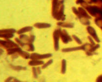 Endospores are tough, highly heat- and chemical-resistant structures
produced
by a few bacterial groups, such as Bacillus and Clostridium, to aid in their
survival. Endospores are always produced inside a bacterial cell and average ~ 1
µm in size (or less). Because of their resistant coat, endospores are difficult
to stain. A special endospore stain (malachite green) can be used but requires
heating to drive the stain into the spores (Atlas
Fig. 4.12). Often, though, spores will be visible after simple staining, as seen
in this photo of Bacillus. Since the rest of the cell is stained, the spore
appears as a circular unstained region within the cytoplasm (Atlas
Fig. 4.13).
Endospores are tough, highly heat- and chemical-resistant structures
produced
by a few bacterial groups, such as Bacillus and Clostridium, to aid in their
survival. Endospores are always produced inside a bacterial cell and average ~ 1
µm in size (or less). Because of their resistant coat, endospores are difficult
to stain. A special endospore stain (malachite green) can be used but requires
heating to drive the stain into the spores (Atlas
Fig. 4.12). Often, though, spores will be visible after simple staining, as seen
in this photo of Bacillus. Since the rest of the cell is stained, the spore
appears as a circular unstained region within the cytoplasm (Atlas
Fig. 4.13).
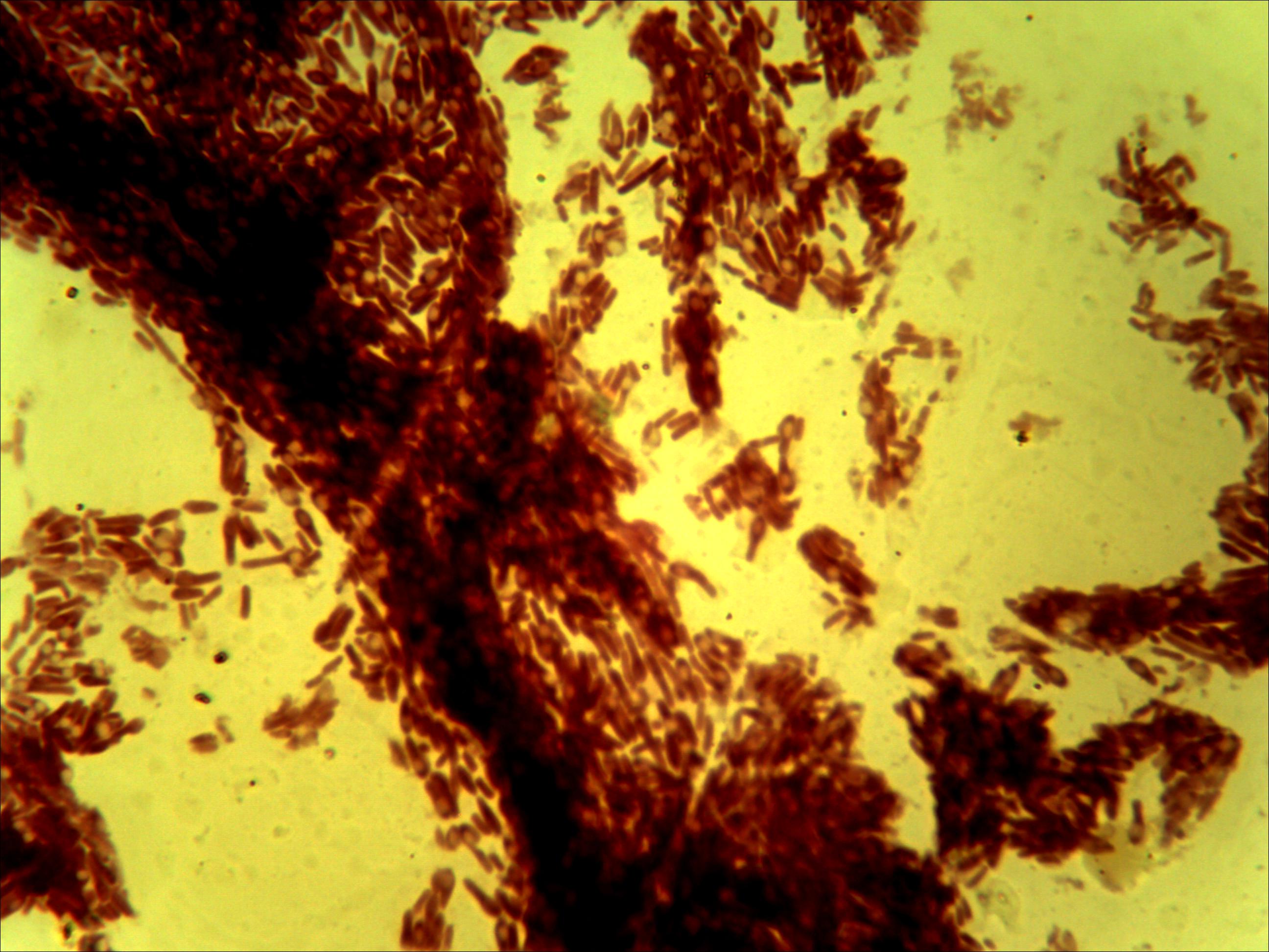
Capsules are protective structures formed
on the outside of a bacterium's cell wall. 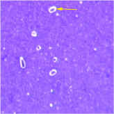 Capsules (sometimes called slime
layers) consist of polysaccharides and/or glycoproteins, which have a slippery
or even sticky mucus-like consistency. Using capsules, bacteria can attach
themselves to surfaces. Streptococcus mutans, the bacterium that causes
tooth decay, uses its capsule to adhere to tooth enamel. For other
bacteria, capsules are a form of protection against the host's immune system. Streptococcus
pneumoniae, for instance, is completely harmless without its capsule, but
causes a deadly form of pneumonia when protected by a capsule. Capsules cannot
be stained easily because of their chemical makeup. Rather, a capsule staining
procedure stains the background (this is known as a negative stain) and, using a
second dye such as safranin, stains the cytoplasm. The only part of the slide
NOT stained is the capsule, now visible as a white halo surrounding the bacteria
(Atlas Fig.
4.10).
Capsules (sometimes called slime
layers) consist of polysaccharides and/or glycoproteins, which have a slippery
or even sticky mucus-like consistency. Using capsules, bacteria can attach
themselves to surfaces. Streptococcus mutans, the bacterium that causes
tooth decay, uses its capsule to adhere to tooth enamel. For other
bacteria, capsules are a form of protection against the host's immune system. Streptococcus
pneumoniae, for instance, is completely harmless without its capsule, but
causes a deadly form of pneumonia when protected by a capsule. Capsules cannot
be stained easily because of their chemical makeup. Rather, a capsule staining
procedure stains the background (this is known as a negative stain) and, using a
second dye such as safranin, stains the cytoplasm. The only part of the slide
NOT stained is the capsule, now visible as a white halo surrounding the bacteria
(Atlas Fig.
4.10).
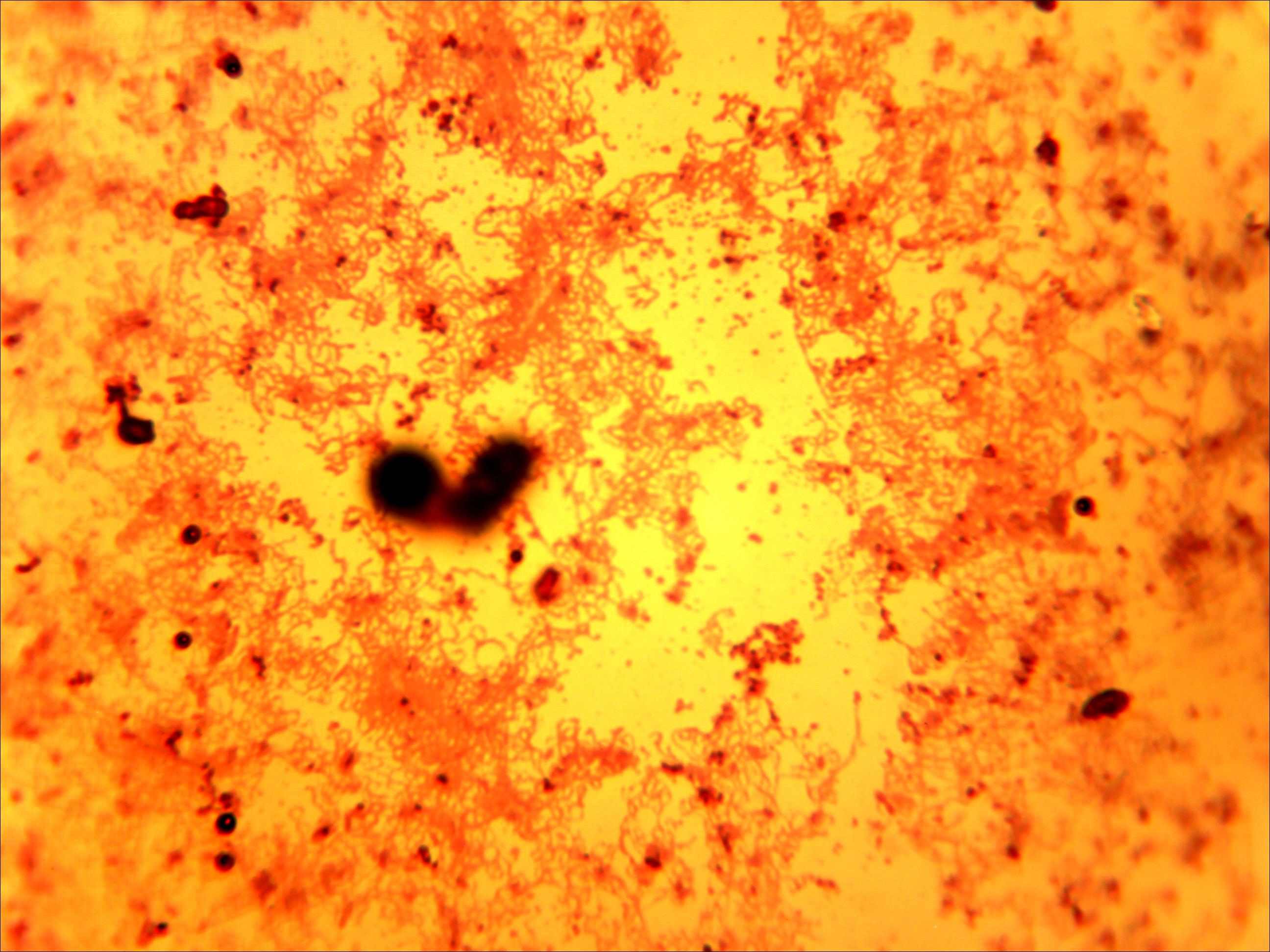
Flagella are long thin appendages which
bacteria use for motility. Composed of the 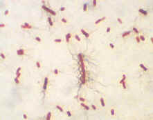 protein
flagellin, flagella are so
thin that they cannot be seen with a light microscope without a special stain.
In a flagellar stain, various compounds (also known as mordants) are used to
attach themselves to the flagella, making them appear thicker and become
visible. Bacterial flagella stains often make the flagella appear more prominent
than they are in actuality. On E. coli, flagella will appear as numerous
squiggly lines surrounding the cell (compare to Atlas
Fig. 4.19). Such an arrangement of flagella is known as
peritrichous (flagella all over the cell surface). Other flagella
arrangements include lophotrichous,
amphitrichous , and monotrichous.
protein
flagellin, flagella are so
thin that they cannot be seen with a light microscope without a special stain.
In a flagellar stain, various compounds (also known as mordants) are used to
attach themselves to the flagella, making them appear thicker and become
visible. Bacterial flagella stains often make the flagella appear more prominent
than they are in actuality. On E. coli, flagella will appear as numerous
squiggly lines surrounding the cell (compare to Atlas
Fig. 4.19). Such an arrangement of flagella is known as
peritrichous (flagella all over the cell surface). Other flagella
arrangements include lophotrichous,
amphitrichous , and monotrichous.
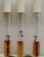
Flagella are the main way by which bacteria accomplish motility, the other (less common) method being gliding motility. bacterial motility can be observed by several means, including the dark-field microscope and a motility test medium. A SIM test can also tell motility in many cases, but because of its yellow color it may be hard to observe.
© 2003 - 2015 José de Ondarza, Ph.D.