Specimen
collection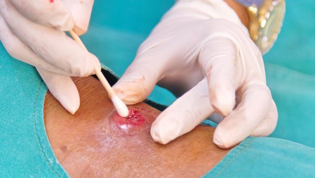
-
Sterile swabs
Specimen handling & transport
-
Labeling
-
Preservation
-
Storage & shipping
Culturing the pathogen
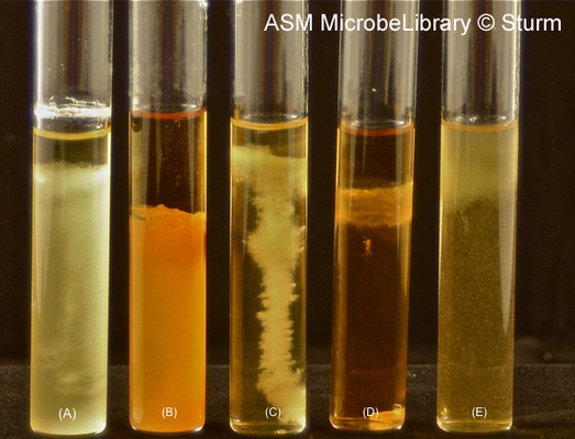
aerobic vs. anaerobic media
-
Thioglycollate medium for Clostridium
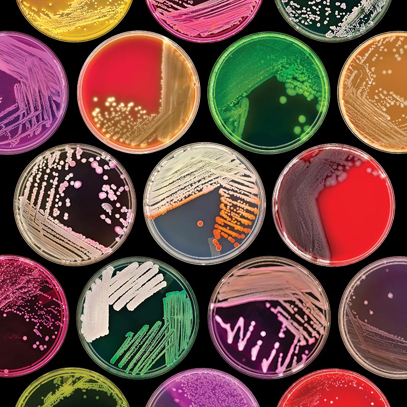
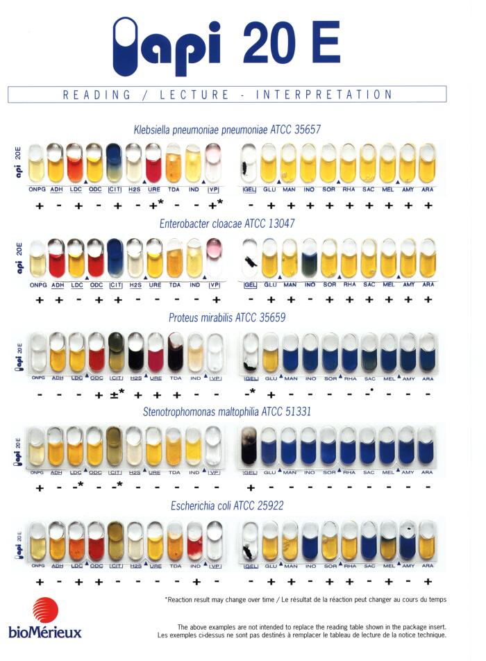
selective and differential media: many types
-
Blood agar: differential for Streptococcus, Staphylococcus
-
Eosin Methylene Blue agar: s/d for enteric bacteria
-
MacConkey agar (s/d for enterobacteria)
-
Thayer Martin agar: s/d for Neisseria, Francisella
-
Mannitol Salt agar: s/d for Staphylococcus
-
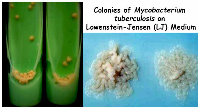
Lowenstein-Jensen medium for Mycobacterium
-
Salmonella-Shigella (XLD) agar - selective/differential
-
CHROMagar- differential for Candida spp., other microbes
-
Sabouraud dextrose agar: fungi
enriched media for fastidious microbes
-
Blood agar: Streptococcus, Bordetella
-
Chocolate agar: Neisseria, Haemophilus
cultivation in host cells
-
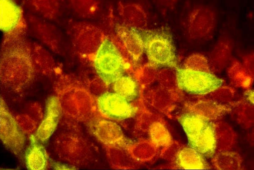
Rickettsia, Chlamydia: cell cultures
-
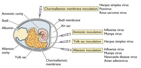
Viruses: cell cultures, chicken eggs
unculturable or impractical-to-culture pathogens
-
Rickettsia: too dangerous
-
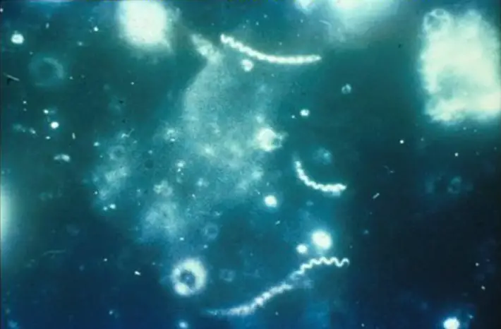
Treponema, Borrelia, Chlamydia, Mycoplasma - difficult to culture
-
Mycobacterium, fungi - grow too slowly
#/media/File:Streptococcal_hemolysis.jpg)